Toll-like receptors - (by Anant Kumar, Replico)
What are Pattern recognition receptors?
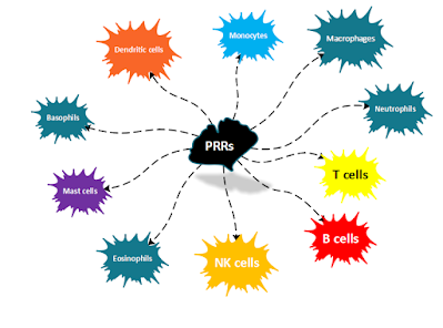
- Pattern recognition receptors are responsible for recognising PAMP on both the extracellular and intracellular pathogens like bacteria, viruses, fungi, parasites, and DAMPs from damaged/dead/dying cells and activation of innate immune responses.
- Expressed on the plasma membrane & inside our cells in lysosomes, endosomes, etc.
- PRRs are also cells that are commonly exposed to infectious agents :
→ mucosal and glandular epithelial cells,
→ vascular endothelial cells that line the blood vessels,
→ fibroblasts and stromal support cells in various tissues.
Toll-like receptors -Discovered in 1996 by Jules Hoff man and Bruno Lemaitre
- Toll-like receptors (TLRs) were the first family of PRRs to be discovered. It is best-characterized in terms of their structure, how they bind PAMPs and activate cells and the extensive and varied set of innate immune responses that they induce. Hoff man and Bruno observed that mutations in toll, made flies highly susceptible to lethal infection with Aspergillus fumigatus, a fungus to which wild-type flies were immune. Later it was observed that Toll and related proteins are involved in the activation of innate immune responses in invertebrates.
- 1997 Charles Janeway and Ruslan Medzhitov à discovered a human gene for a protein similar to Toll that activated the expression of innate immunity genes in human cells. So far, 10 TLRs that function as PRR are identified in humans and 13 in mice.[1] (but humans have 11)
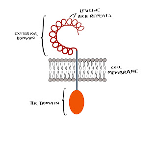
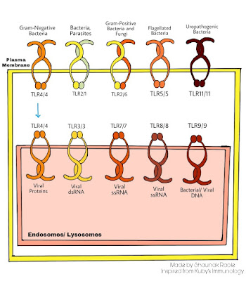
- TLRs are transmembrane proteins. All TLRs have an amino-terminal exterior domain and a carboxy-terminal TIR domain that interacts with TIR containing adapter. The Exterior domain is called leucine-rich repeats (LRRs); multiple LRRs make up the horseshoe-shaped extracellular ligand-binding domain of the TLR polypeptide chain. TLR dimerize to form homodimer or heterodimer(TLR2 forms heterodimers by pairing with either TLR1 or TLR6). It interacts with the PAMP OR DAMP with its dimerized exterior domain.
- TLRs on the plasma membrane recognize PAMP on the outer surface of extracellular microbes. TLRs in endosomes and lysosomes recognize internal microbial components – nucleic acid in particular
24-29 amino acids containing the sequence LxxLxLxx, where L is leucine and x is any amino acid, interior Toll/IL-1R (TIR) domain
- Nucleic acid-sensing TLRs are TLR3, TLR7, TLR8, TLR9 (Blasius and Beutler 2010)TLR4 has been shown to move from the plasma membrane to endosomes/ lysosomes after binding LPS or other PAMPs.In addition to microbial ligands, TLRs also recognize DAMPs, endogenous (self) components released by dead/ dying cells or damaged tissues. Among the DAMPS recognized by plasma membrane TLRs are heat shock and chromatin proteins, fragments of extracellular matrix components (such as fibronectin and hyaluronan), and oxidized low-density lipoprotein and amyloid-β. The cytoplasmic domain of TLR is homologous to those of IL-1R.
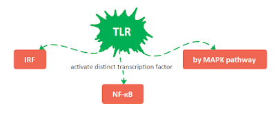
TLRs signalling pathway is a combination of some shared pathways and some TLRs specific pathways.
- Shared pathway → NF-КB activation --> it induces innate and inflammatory genes → encoding defensins; enzymes such as iNOS; chemokines; and cytokines such as the pro-inflammatory cytokines TNF-α, IL-1, and IL-6, produced by macrophages and dendritic cells
- TLR specific pathways → ex. interferon regulatory factors (IRFs) → induce transcription of IFN-α and INF-β
- Signal transduction depends on the TLR & protein adapter which binds to the cytoplasmic domain
- TIR domains of all TLR dimers serve as binding sites for the TIR domains of adaptors that activate the downstream signalling pathways.
- TLRs need to dimerise to initiate the downstream signalling events. ((Gay and Gangloff, 2007))
TLRs that interact with extracellular ligands reside in the plasma membrane; TLRs that bind ligands generated from endocytosed microbes are localized to endosomes and/or lysosomes. Upon ligand binding, the TLR4/4 dimer moves from the plasma membrane to the endosomal/lysosomal compartment, where it can activate different signalling component
→ TLR3, TLR7, and TLR9 are subject to stepwise proteolytic cleavage, required for ligand binding and signalling (Barton and Kagan 2009).
- Whether found on the cell surface or in endosomes/ lysosomes, most TLRs bind the adaptor protein MyD88 (activating MyD88-dependent signalling pathways).
- TLR3 binds the alternative adaptor protein TRIF (activating TRIF dependent signalling pathways).
- TLR4 is unique in binding both MyD88 (when it is in the plasma membrane, signalling its endocytosis) and TRIF (when it is in endosomes, after internalization).
- → Additional adapter à serve as sorting receptors: TIRAP helps to recruit MyD88 to TLRs 2/1, 2/6, and 4, and TRAM helps to recruit TRIF to both TLR3 and endosomal/lysosomal TLR4.
The two key adaptors are MyD88 (Myeloid differentiation factor 88) and TRIF (TIR-domain-containing adaptor-inducing IFN-β factor).
MyD88-Dependent Signalling Pathways 1.1
- MyD88 initiates a signalling pathway that activates the NF-кB
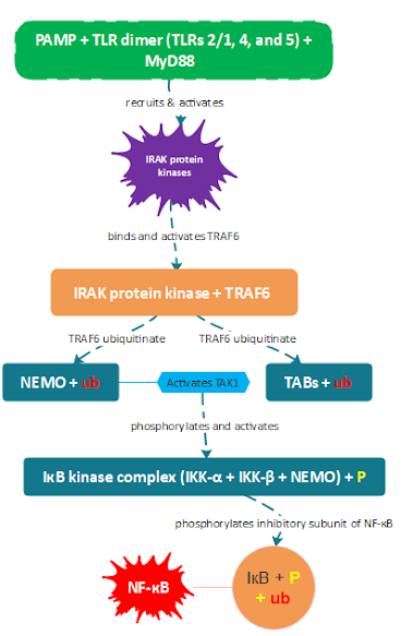
- TLRs 2/1, 4, and 5, MyD88 recruits and activates several IRAK protein kinases, which then bind and activate TRAF6. TRAF6 ubiquitinates NEMO and TAB proteins, leading to the activation of TAK1, which then phosphorylates the IкB kinase (IKK) complex.
- Activated IKK then phosphorylates the inhibitory IкB subunit of NF-кB, releasing NF-кB to enter the nucleus and activate gene expression.
1.2
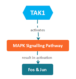
→ After separating from the IKK complex, TAK1 activates MAPK signalling pathways that result in the activation of transcription factors including Fos and Jun, which make up the AP-1
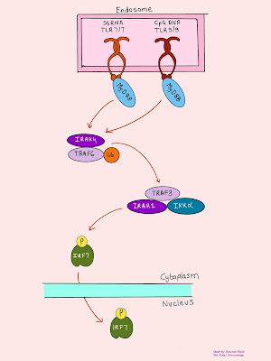
→ the endosomal TLRs 7, 8, and 9, bind microbial nucleic acids
→ viral RNA and
→ bacterial DNA
→ MyD88/IRAK4/TRAF6 complex activates a complex containing TRAF3, IRAK1, and IKKα, à leading to the phosphorylation, dimerization, activation, and nuclear localization of IRF7.
→ IRF7 induces the transcription of genes for both Type 1 interferons, IFN-α and -β, which have potent antiviral activity.
TRIF Dependent Signalling Pathway
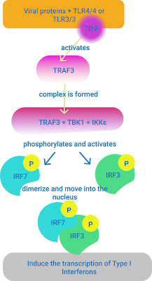
→ TRIF activates TRAF3
→ IRF3 and IRF7 dimerize and move into the nucleus and (together with NF-кB and AP-1) induce the transcription of the IFN-β and -α genes
intracellular TLRs that bind viral PAMPs → induce the synthesis and secretion of Type I interferons → inhibit the replication of the virus in that infected cell.
→ TRIF recruits the RIP1 protein kinase that in turn recruits and activates TRAF6, which then enters the 1.3 pathway
[1]Lim, K. H., & Staudt, L. M. (2013). Toll-like receptor signalling. Cold Spring Harbor perspectives in biology, 5(1), a011247. https://doi.org/10.1101/cshperspect.a011247
[2]Anthoney, N., Foldi, I., & Hidalgo, A. (2018). Toll and Toll-like receptor signalling in development. Development (Cambridge, England), 145(9), dev156018. https://doi.org/10.1242/dev.156018
[3]Kuby Immunology 7th Edition
Very informative 🔥
ReplyDeleteThank You sam
DeleteGood work 👌
ReplyDeleteGood work bro👌
ReplyDeleteGreat info
ReplyDeleteNice work 👍👍
Thank you
DeleteIncredible information in this tiny blog ☺️☺️
ReplyDeleteNice work
ReplyDeleteGreat work bro 👍
ReplyDelete