Immunotherapy for Cancer
Immunotherapy for Cancer (by Shaunak Raole, Replico)
Immunotherapy, also referred to as biological therapy, is done to treat a disease either by activation/enhancement or suppression of the immune system. When the immune system is amplified, it is called activation immunotherapy, and when it is suppressed, it is called suppression immunotherapy.
Inactive
immunotherapy, the immune response against cancer is triggered and in passive
immunotherapy, immunity molecules are administered to patients who can not
produce them on their own.
A lot of
potential has been seen in immunotherapy, especially against cancer.
Immunotherapies
have been designed to eliminate a tumour by reviving, initiating or
supplementing the in vivo anti-tumour immune response. They can also neutralize
the inhibitory pathways.
There are
many classes of immunotherapy:
Monoclonal Antibodies against Tumour Cells
The immune system produces antibodies when it senses a threat. These antibodies are
proteins that interact with antigens and lead to an immune response.
Monoclonal antibodies are produced in labs and boost the natural antibodies or
defend against foreign threats.
The mAbs can
be unmodified or they can be conjugated with some agent to improve their
efficacy. For example, potent toxins, chemical agents, chemotherapy drugs, or
radioactive particles can be conjugated with a mAb which then can be delivered to
the target site/cell. These are also called “guided missile therapies”. In this, the toxic agents are specifically delivered to the tumour cells. The normal
tissues are spared.
Certain
reagents known as immunotoxins have been synthesised. This is done by coupling
the inhibitor chain of a toxin like Diptheria toxin to an antibody against a
tumour-specific or tumour associated antigen.
Some tumours express significantly high levels of growth factors which can be promising
targets for anti-tumour mAbs. As an example, in 25% to 30% of women having
metastatic breast cancer, there is a genetic alteration of tumour cells which
leads to increased expression of human epidermal-growth-factor-like receptor 2
(HER2), encoded by the neu gene, and is present in trace amounts in normal
adults. Thus, a humanized mAb against HER2 has been successfully used to treat
breast cancers in which HER2 is expressed. Normal cells aren’t damaged in the
process.
Bispecific
monoclonal antibodies (BsmAb) are antibodies that bind with two proteins at
once, and some can bind to both cancer cells and an immune system cell, which
facilitates the immune response against cancer.
Approximately
12 different monoclonal antibodies have been approved for treating cancer.
Checkpoint Inhibitors
This is the most thoroughly investigated class of immunotherapy, in which the two most common strategies are: PD-1/PD-L1 blockade and CTLA4 inhibition. Certain immune checkpoints are responsible for maintaining appropriate immune responses and protecting healthy tissues from immune attacks. So these are basically molecules present on specific immune cells and need to be either activated or inactivated for the immune response to get elicited.
When T cells activate in response to inflammation, PD-1, a checkpoint protein, is expressed by them which allows them to recognize abnormal and cancerous cells. It also prevents the T cells from attacking other cells in the body. The prevention occurs when the cells express PD-L1 protein. This is because if PD-1 binds to PD-L1, T cells are stopped from attacking any cell. The tumour cells express PD-L1 which binds to PD-1 and renders T-cells inactive. If we use mAbs that target PD-1 or PD-L1, this interaction can be blocked and tumour cells can be killed.
Some drugs which target PD-1/PD-1 inhibitors: Pembrolizumab (Keytruda), Nivolumab (Opdivo), cemiplimab (Libtayo). Drugs that target PD-L1 (PD-L1 inhibitors) are Atezolizumab (Tecentriq), Avelumab (Bavencio), Durvalumab (Imfinzi).
CTLA-4 (cytotoxic T-lymphocyte-associated protein 4), which is a co-inhibitory molecule regulates the degree of T-cell activity and is another immune checkpoint. T cell activity gets inhibited by the interaction between CTLA4 and its ligands, CD80 and CD86. This can promote tumour progression. Hence if we are able to block that interaction, T cells can remain active and recognize and kill the tumour cells. Unfortunately, the exact mechanism of the CTLA4 blockade is unknown and the various antibodies which target CTLA4 have different properties. For example, some anti-CTLA4 antibodies inhibit checkpoint functionality and also deplete regulatory T cells. Ipilimumab (Yervoy) is a CTLA4 targeting mAb.
The mAbs which target these checkpoints have been FDA-approved for the treating malignant cancers like melanoma, non-small cell lung cancer, RCC, Hodgkin lymphoma, Merkel cell carcinoma, head and neck cancer and carcinoma of the bladder

Cytokines
IFN α therapies were approved in 1986, hence cytokines were the first class of clinically applied immunotherapy. Cytokines are injected and directly stimulate the growth and activity of immune cells.
Interferons
(IFN-α, -β, and -ϒ), Interleukins (IL-2, -4, -6, and -12) and
granulocyte-macrophage colony-stimulating factor (GM-CSF) are the three main
types of cytokines used for immunotherapy. The immune cells normally
produce interferons as a response against microbial pathogens. Interferons elicit an immune response by inducing the maturation of macrophages, NK cells, lymphocytes
and dendritic cells. Angiogenesis in the extracellular tumour space can also be
inhibited by interferon activation.
The activity
and growth of CD4+ T cells and CD8+ T cells, along with NK cells, can be
stimulated by Interleukins, mainly Interleukin 2. Activated myeloid cells
produce IL-15 as a membrane-bound heterodimer (IL-15Rα associated) such that it is trans-presented to
NK cells and T cells express IL-2/IL-15Rβ and the common ϒ chain receptor. IL-15
has critical importance for the ontogeny of NK cells and CD8+ T cells. It induces
proliferation, cytotoxic action and release of other cytokines like IFN-gamma
from these cells, which indicates its role in potentiating immune response.
GM-CSF is
responsible for improving immune response by promoting T cell homeostasis
(which improves T-cell survival) and by supporting dendritic cell
differentiation in order for these cells to express tumour specific antigens.
Granulocyte
colony-stimulating factor (G-CSF) and GM-CSF have both been used to augment and
accelerate granulocyte recovery after chemotherapy, however, GM-CSF is more
pro-inflammatory.
Along with
these cytokines, certain agonists are being investigated which activate immune
cells via intracellular mechanisms. As an example, TGFβ receptor type 1 (TGFβR1) inhibitors like SD-208 restore
functions of T cells and bring an improvement in the immune response. APCs are directly
activated by the small molecule agonists of TLR7/TLR8 to promote antitumour
activity. STING (Stimulator of interferon genes) agonists induce
pro-inflammatory cytokine production and other types I interferon responses.
Engineered T cells: chimeric antigen receptor T and T cell receptor T
cells.
The extracellular domain of CAR
contains the antigen-binding moiety along with a spacer. The antigen-binding
moieties can either be an scFv (single-chain fragment variable) which is derived
from antibodies, or a human Fab fragment taken from phage display libraries, or
nature ligands that engage their cognate receptor.
The scFv is a fragment derived from
mouse mAbs humanized mAbs or human mAbs and is responsible for the recognition
and binding to the tumour associated antigens (TAAs) which are expressed in the
tumour cell surface.
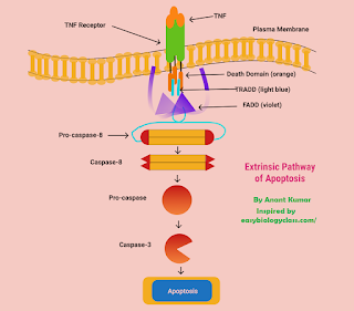
Along with unprocessed antigens, these receptors can also recognize carbohydrate and glycolipid structures present on the tumour cell surfaces. Antigen presentation via MHC is bypassed. The CAR T-cells (CD4+ and CD8+) are recruited for redirected recognition of target cell, and CAR mediated tumour elimination uses at least two pathways in executing cytolysis, for example, perforin and granzyme exocytosis. Death receptor signalling via Fas/Fas-ligand or TNF/TNF-receptor can also take part to some extent.
These CAR T cells retain their activity for over 10 years after injection, hence this can be called a one-time therapy. These cells are bring clinically tested for haematological as well as solid cancers.
Engineered TCR T cells are currently
undergoing clinical trials for haematological and solid cancers. The TCRs
interact with tumour-associated intracellular antigens which are presented by
MHCs. The antigenic target can be shared antigens like cancer-testis antigens,
or patient-specific neoantigens arising from tumour mutations. Contrary to MHC
independent CAR T Cells, the TCR T cells need to be MHC matched with the
patient.
As of now, researchers are developing novel
delivery technologies for overcoming toxicities associated with CAR T Cells and
TCR T cells, and to increase their applicability for solid tumours.
Co-stimulatory receptor agonists
Certain agonistic antibodies can be designed to bind specifically to receptors present on T cell surfaces and trigger intracellular signalling pathways which induce T cell growth, survival and effector functions against tumour cells. The most common of these receptors are the co-stimulatory receptors (namely CD28) and many members of the tumour necrosis factor receptor (TNFR) family like 4-1BB (also called TNFRSF0 or CD137), OX40 (also called TNFRSF4) and glucocorticoid-induced TNFR-related protein (GITR) which are expressed on APCs surfaces. The binding of ligands to these co-stimulatory receptors trigger intracellular cell signalling which in turn promotes T cell growth and anticancer activity.
Since agonistic antibodies are at the early development stage, there aren’t any approved by the FDA. However, several
have reached clinical trials.
Cancer Vaccines
Also called therapeutic vaccines, they
are given to people already suffering from cancer in order to increase the
body’s natural defence. They either prevent cancer from recurring, or they
destroy remnant cancer cells (from other treatments). They can also stop a
tumour from spreading.
There are various types of cancer
vaccines. They include tumour cell lysate, dendritic cells, nucleic acids (like
mRNA) or neo-antigens. The most commonly studied class of cell-based cancer
vaccines are dendritic vaccines. They are made up of dendritic cells which are
collected from patients and are engineered such that they express tumour
associated antigens and hence directly activate T cells to attack tumour cells.
Sipuleucel-T is the first Dendritic Cell-based cancer vaccine for
asymptomatic/minimally symptomatic metastatic castration-resistant prostate
cancer. It induces an immune response to a common self prostate tumour
antigen, prostatic acid phosphatase (PAP). The DCS is isolated from prostate
cancer patients. Next, they are stimulated in vitro using a fusion protein that
consists of PAP and GM-CSF. The expanded autologous APCs are then re-infused
into the patient yielding an increased survival of just over 4 months.
Nucleic acid therapeutics (DNA and RNA
based vaccines) are good alternatives to conventional vaccines. They rely on
intracellular delivery of exogenous nucleic acids into the target cells. APCs
take up DNA or mRNA, which are translated for inducing antigen expression. T
cells, after being presented with the targeted antigens, are then activated
against tumour cells that express antigen of interest. DNA vaccines are
sometimes unsuccessful due to nuclear delivery barriers and immunogenicity;
hence mRNA cancer vaccines are developed.
Neoantigen vaccines have the ability
to boost immune responses against cancer cells. Neoantigens are specific to
tumours and arise from somatic DNA alterations in cancer cells. The biggest
advantage of these is that they are only present on cancer cells, hence
off-target adverse effects are absent. Neoantigen vaccines are ideal for treating
heterogenous cancers as the vaccines can encompass an unlimited number of
neoantigens.
Oncolytic virus therapy
In this, viruses are modified in the
laboratory such that they infect and kill specific tumour cells. The procedure
is as follows:
- The virus is genetically modified and injected into the tumour.
- It reaches the cancer cells and makes a copy of itself.
- The cancer cells are disrupted and die.
· Once the cells die, the immune system is
stimulated to attack any cancer cells in the body having similar proteins as
the dead cells. When the infected cancer cells are destroyed by oncolysis, they
release virions that help in destroying the remaining tumour.
This oncolytic virus does not affect
any healthy cells. Since it does not depend on any specific antigen expression
patterns, it is considered as superior immunotherapy. Some features which
make it the ideal candidate for treating diverse malignancies are:
· -They enhance the recruitment of
tumour-infiltrating lymphocytes (TILs).
-Reprogramming of immunosuppressive tumour
microenvironment (TME).
-Boost systemic anti-tumour immunity.
The viruses have two kinds of
interactions with the immune system.
1. Immunity
as an obstacle
The immune system naturally attempts to deactivate any virus. Hence, immunosuppression by chemotherapy and inhibition of the complement system has to be done. Since antibody generation cannot be avoided, the viral vector can be coated with polyethene glycol to shield it from antibodies. The viruses can also be hidden inside macrophages, as they automatically migrate towards areas of tissue destruction and where oxygen levels are low. Such sites are characteristic of cancer growth.
2. Immunity
as an ally
The infection of the virus can attract the attention of the immune system to the tumour and can help in the generation of useful and long-lasting antitumour immunity. This can also allow the production of a personalized cancer vaccine. Some immunogenic oncolytic viruses, after infecting the tumour cells, can elicit an anti-tumour immune response. This is especially true in the case of viruses delivering cytokines or other immune-stimulating factors. Imlygic is an attenuated herpes simplex virus that has been engineered genetically to replicate preferentially within tumour cells and generate antigens that can trigger an immune response.
Genetically unmodified ECHO-7 strain enterovirus RIGVIR was the first oncolytic virus to be approved by a national regulatory agency for treating melanoma.
Using Immunotherapy for other conditions
· Guillain-Barre syndrome is a common
cause of acute neuromuscular weakness and paralysis around the world.
Intravenous immunoglobulin (IVIg) and plasma exchange (PE) are methods of
immunotherapy for GBS.
· Food Allergy is a serious pathological cascade of immune responses to molecules or molecular fragments.
Oral immunotherapy can be used to redirect the atopic immune responses to food
allergy patients, as the patients ingest small and gradually increasing
allergen doses over some months, triggering safer immune responses against
these antigens.
· Covid 19 has become a global threat and deaths keep increasing every day. An immunotherapeutic agent can be developed which targets the neutralizing epitopes in the RBD of the virus. This is called immunofocusing.
by Shaunak Raole
https://replicoo.blogspot.com/2021/07/toll-like-receptors.html
https://replicoo.blogspot.com/2021/06/microbial-bioinformatics-game-changing.html
https://replicoo.blogspot.com/2021/07/spyder-python-install.html

Nice Work Shaunak
ReplyDeleteThank you!
DeleteVery well articulated
ReplyDeleteThank you!
DeleteGreat work 🔥
ReplyDeleteThank you!
DeleteWow
ReplyDeleteThank you!
DeleteAmazing dawg🙌🏼
ReplyDeleteThank you!
DeleteThis blog was really very informative and even though I am from a non-bio background, I understood it perfectly!
ReplyDeleteI'm glad, thank you!
Delete